Every minute, 250 infants are born worldwide, and two of those are born with a congenital heart defect. Severe cases require immediate surgical intervention, and for a pediatric cardiac surgeon, early and accurate detection is essential to creating a successful patient care plan.
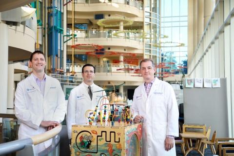
In the Division of Cardiovascular and Thoracic Surgery, Andrew Lodge, MD, Joseph Turek, MD, PhD, and Nicholas Andersen, MD, work closely as a three-person surgical team, part of a larger interdisciplinary pediatric cardiac program at Duke.
A High Wire Act
Dr. Lodge, Associate Professor of Surgery and Pediatrics and member of the division since 2003, worked for years to help build the program and provide clinical care to an ever-growing number of patients. It was a challenging task, as he was the only congenital heart surgeon at Duke for two different periods.
“When I started 16 years ago, the program was smaller and my responsibilities were split between congenital heart surgery and adult heart transplant and ventricular assist
devices,” says Dr. Lodge. “About 8 or 9 years ago, I became the sole congenital heart surgeon for a period of about 6 months. At the time we had a productive lab, but not as much protected time for research, and as we were growing clinically it didn’t leave a lot of time for non-clinical pursuits.”
Changes came in 2014, when Allan D. Kirk, MD, PhD, Vice Dean of the Section of Surgical Sciences, assumed the role of department chair and began to allocate resources for program growth.
“Dr. Kirk made a commitment to fully staff each section in children’s surgery with a minimum of three people to manage the clinical enterprise,” says Dr. Lodge. “With the additional manpower, we could be academically productive, or productive in other pursuits. This commitment allowed us to add Joe [Turek] and Nick [Andersen] to the team.”
When Dr. Turek, Associate Professor of Surgery and Pediatrics and Chief of the Section of Pediatric Cardiac Surgery, joined in 2017, he collaborated with Dr. Lodge to improve care for patients with congenital heart disease.
“When I arrived, we worked on some changes to our programmatic approach,” says Dr. Turek. “Pediatric heart surgery is not just about the operation. It’s not just about the preoperative care or postoperative care. It is a fine balance between all of these things. It’s a high wire act—everyone has to be performing well.”
A Game of Millimeters
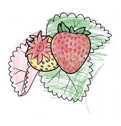
For the surgical team, achieving this balance starts with an adequate preoperative strategy. Preparation for surgery on a heart sometimes no larger than a strawberry presents unique challenges. Dr. Andersen, Assistant Professor of Surgery, joined the team in 2018 and speaks to the delicate nature of surgery on children.
“There is no margin for error,” he says. “Our field is a game of millimeters, and sometimes a one-millimeter difference can mean life or death for an infant or child.”
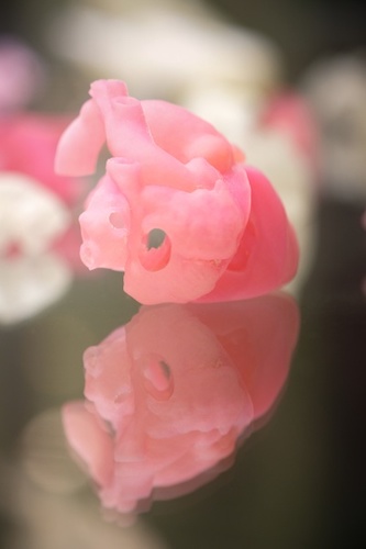
Most preparatory imaging modalities, such as echocardiograms, CT scans, or MRIs, are valuable tools but offer a limited perspective: a two-dimensional picture that must be mentally converted to analyze spatial relationships. Through a partnership with the Division of Cardiology in the Department of Medicine, the team uses flat images to create 3D-printed models, valuable assets in charting an appropriate surgical course.
“The 3D model is as good as it gets,” Dr. Andersen says, “to examine how a pathway between chambers connects, to assess if there are any obstructing muscle bundles you have to work around. You can plan essentially where you are going to put every single stitch of the repair.”
The models also help in decision-making as to whether an invasive or non-invasive procedure is necessary.
“The 3D models give us an idea of whether the heart is amenable to putting a stent in,” Dr. Turek says. “For babies who have insufficient blood flow to the lungs, we primarily like to stent patients in the catheterization lab now, if at all possible. If not, we then move toward doing a surgically placed shunt.”
The Room Where It Happens
Best laid plans are only as good as the surgeons performing in the operating room, and there, two sets of eyes are better than one.
Teaming up for surgeries has been an effective programmatic change. Using a 1–5 rating system, high-profile, complex cases in the 4–5 range are flagged, requiring two attending surgeons.
“Having two fully trained, board-certified surgeons has made a difference,” says Dr. Andersen. “Our outcomes are heavily scrutinized and gain lot of attention in national press. With two of us in the OR, we can perform faster, safer, and with far better results.”
“Complex cases are where you incur higher mortality,” adds Dr. Turek. “To make this work, we double-scrub on a lot of cases and it is a huge time commitment, but it has greatly improved quality.”
As case numbers rise, so does the incidence of cases that require highly-specialized skills for effective treatment. Single ventricle defects, which result in an underdeveloped left side of the heart, are considered by the surgical team to be the last frontier of pediatric heart disease.
The Norwood Procedure, a common but risky operation for single ventricle defects, is one that Dr. Turek has focused on improving due to its 15–20 percent national mortality rate.
“The Norwood Procedure is the highest risk thing we do,” he says. “I’ve developed the operation so that we perform it while giving blood flow to the entire body. For these cases we use a heart–lung machine, and we work with the perfusionists to make sure we are getting proper flow to the proper locations.”
In their recent series of Norwood operations employing this all-region perfusion strategy, mortality has decreased to less than 10 percent.
When Repair Isn’t an Option
For congenital defects that are too severe for repair, the last resort is heart transplant.
Even after a successful transplant, the patient’s heart needs to be replaced, on average, every 14 years. Life for a child after a heart transplant means potentially undergoing several more highly invasive surgeries. A solution for developing tolerance to the transplanted heart could mean fewer future transplants and better long-term patient health.
But how? The immune system is complex and often works in nebulous ways. A solution could be found in the thymus, which trains its T cells to create an ample defense system in recognizing self and non-self proteins. By co-transplanting this captain of the immune system with another organ, you could theoretically retrain the recipient’s immune system and steer the ship in a new direction. Duke, it turns out, is uniquely suited for this type of research.
Duke is the only center in the Western Hemisphere that performs thymus transplants, a specialty that originated at Duke 25 years ago. Professor of Pediatrics Mary Louise Markert, the impetus of the thymus transplant program, began studies in 2012 in an animal model to test thymus transplant as a way to induce tolerance.
“We’ve been talking about co-transplant for years with Dr. Markert,” Dr. Lodge says. “She really thinks outside the box, and one of the ideas she shared was the tolerance-induction model. Our group didn’t at first have the resources to pursue it, but now we can.”
With the team’s expansion allowing protected time for research, Dr. Turek has worked with Dr. Markert to continue this project.
“We’ve successfully demonstrated tolerance in a rat model,” Dr. Turek says. “We’ve received a cooperation grant and applied for a DOD grant to continue this study in non-human primates. We’re funded to continue these studies there, with an eye on introducing this groundbreaking work in infants in the near future.”
If successful, this research has the potential to improve tolerance in other types of transplants in the future.
"Based upon our programmatic growth in 2019, we look to be in one of the top 10 programs in the country according to volume this year."
- Joseph Turek, MD, PhD
Continued Growth
Though still evolving, the congenital heart surgery program has seen measurable differences in only a few years. Even with an increase in patient volume and a higher case mix index, outcomes have improved.
“2019 for us has been such a productive period,” says Dr. Turek. “We have less than one-half percent mortality for all of our pediatric congenital heart cases. On top of that, the program has grown incredibly. Based upon our programmatic growth in 2019, we look to be in one of the top 10 programs in the country according to volume this year.”
Dr. Lodge says that the team’s success relies heavily on the complementary nature of their different skillsets.
“We have skillsets that overlap to some extent, but also complement each other,” he says. “My strongest skillset is in the operating room and the ICU, so I can provide a lot of perspective and guidance clinically. Joe is great at focusing on program growth and expansion, with an innovative side and new approaches to things. Nick has a strong academic background as a resident and fellow, and he can continue that research here at Duke.”
As the program continues to grow and mature through the team’s efforts, the children under their care are given a much stronger chance of doing the same.