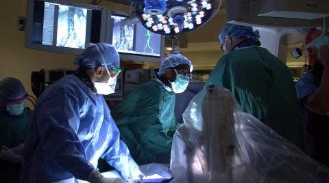
Photo: Dr. Kevin Southerland, General Surgery Chief Resident, inserts a stent graft using CYDAR imaging
In April, Duke vascular surgeons introduced novel 3D imaging that harnesses the power of the cloud to facilitate endovascular aneurysm repair (EVAR).
Developed by CYDAR Medical in the UK, fusion imaging combines cloud computing with 3D overlay technology, producing an on-screen image that “fuses” computed tomography (CT) images taken before surgery with live fluoroscopic images during the procedure, giving surgeons a visual roadmap to help guide stent graft insertion.
Because this technology is connected to the cloud, the overlays update in real time, providing surgeons with a highly accurate image of the patient’s vascular anatomy even if the patient moves. Surgeons can pinpoint blood vessels for inserting guide wires with an accuracy better than 1/5 of a millimeter.
“This technology allows the CT imaging to be superimposed on the fluoroscopic image and it allows the surgeon to see the image in 3D so that when he or she is trying to do a manipulation it’s very much faster,” says Dr. Cynthia Shortell, Chief of Vascular and Endovascular Surgery. “Any time you can reduce operative time, it’s safer for the patient.”
CYDAR imaging has reduced operative time for EVAR cases by approximately one hour, says Dr. Shortell. This shorter time in the OR means a lower amount of radiation during the procedure and a decreased risk of radiation exposure for both the patient and OR staff. Radiation can cause severe complications, including cancer, eye damage, infertility, and radiation burns. In a recent clinical trial, CYDAR imaging reduced X-ray screening time by almost 35%.
Compared with other imaging modalities, CYDAR imaging requires a smaller amount of iodinated contrast during fluoroscopy. Contrast can damage the kidneys and lead to renal insufficiency in patients, known as contrast-induced nephropathy. Recent findings from the CYDAR Infrarenal Endovascular Aneurysm Repair trial indicated a 50% reduction in contrast during fluoroscopy.
Dr. Shortell says the more complex the procedure, the more benefit to the patient. CYDAR imaging is especially helpful in cases that require a custom-made fenestrated stent graft with holes for the renal arteries.
In addition to improving patient safety, this faster procedure time means a shorter hospital stay and reduced costs for patients.
Dr. Shortell envisions wider applications of the technology for any procedure that uses 3D imaging.
“We will use CYDAR imaging in our other hybrid OR for patients undergoing heart surgery. This technology can theoretically be used for any sort of procedure that is done in 3D, including treatments for cancer, so it doesn’t have to be limited to blood vessels.”