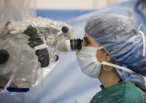
The Duke Department of Surgery launched its new Microsurgery Core with Dr. Linda Cendales serving as core director. Microsurgery uses high-powered microscopes to assist in the surgical repair of tissues, including small vessels and nerves. This technique is utilized in many specialties, such as general surgery, ophthalmology, gynecology, urology, orthopedic surgery, neurosurgery, oral and maxillofacial surgery, plastic surgery, and pediatric surgery.
The Core for Microsurgery and Surgical Models in Small Animals is a multi-dimensional, research-oriented program that provides space and resources for investigators to design models requiring microsurgery and to effectively test their hypotheses. Currently, models are available in kidney, liver, intestinal, hindlimb, abdominal wall, aorta, lung, and heart transplantation. Core members provide technical and operational support and collaborate with investigators to develop advanced models and practices in microsurgery.
The core has various services to assist in the learning, experimental, and surgical practice phases of microsurgery. The Microsurgical Skills and Techniques Training Course offers training in the use of the operating microscope and associated microsurgical instruments, basic microsurgical dissection, and suturing techniques. The Core also offers training in procedures, including artery and vein anastomoses [end-to-end and end-to-side repair], interpositional grafts, tubal repair, vasovasostomy repair, neurorrhaphy, and free tissue transfers.
The Microsurgery Core immerses investigators in a realm of innovative microsurgical design and practice techniques, which may be used in a variety of surgical specialties.